


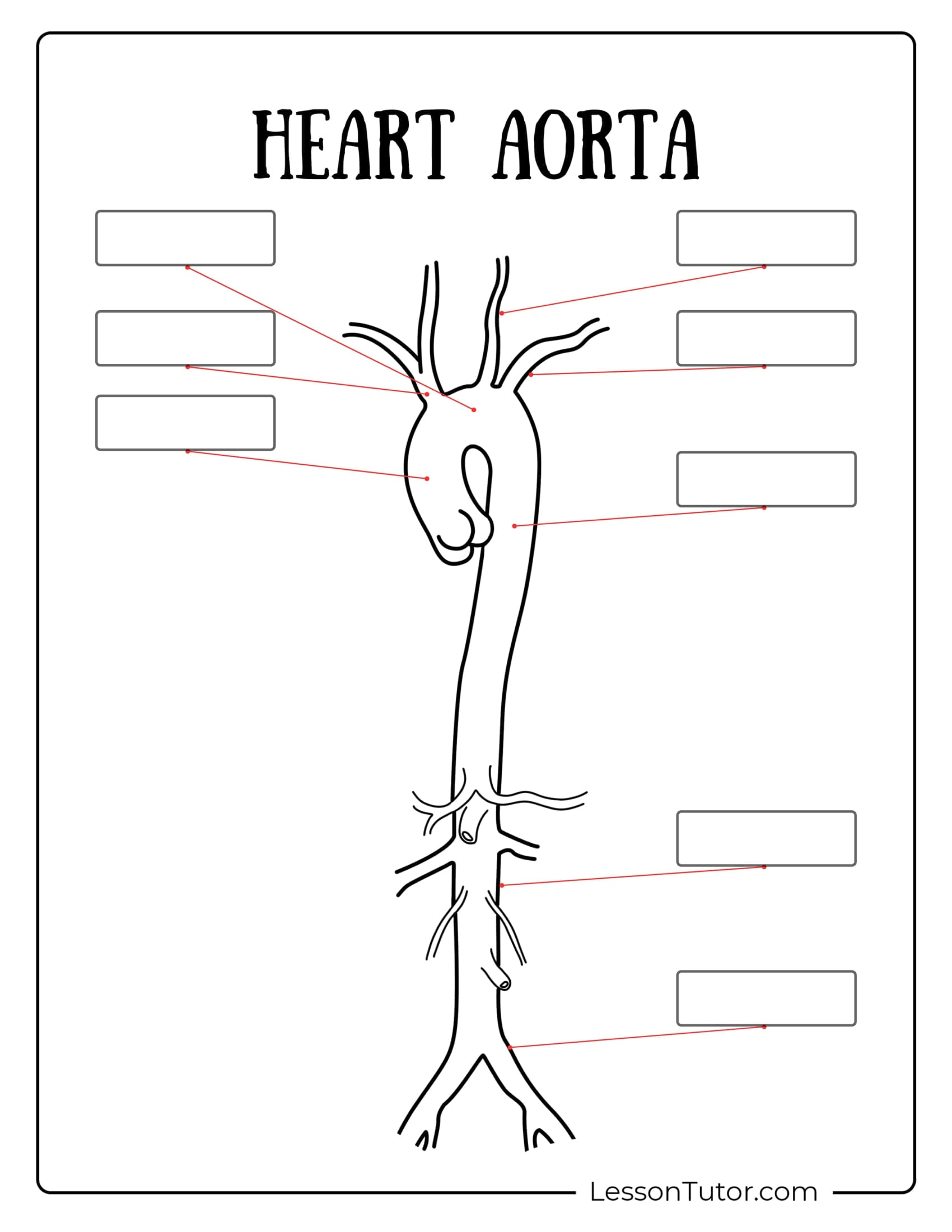
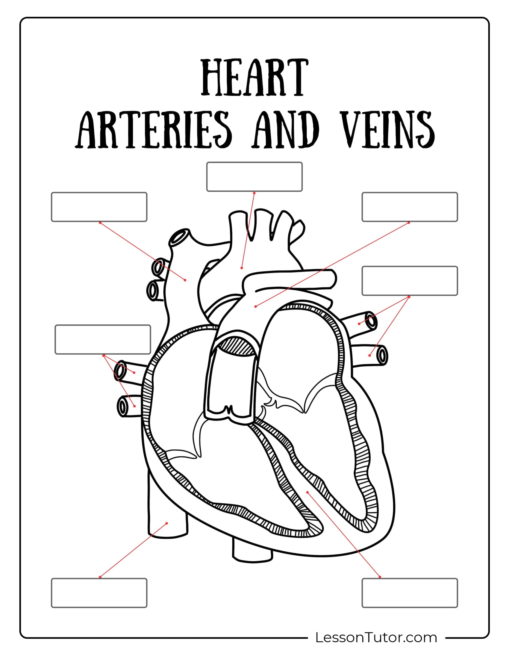
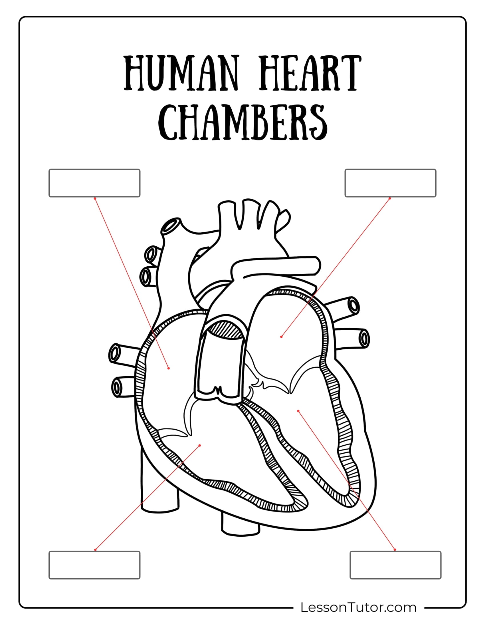
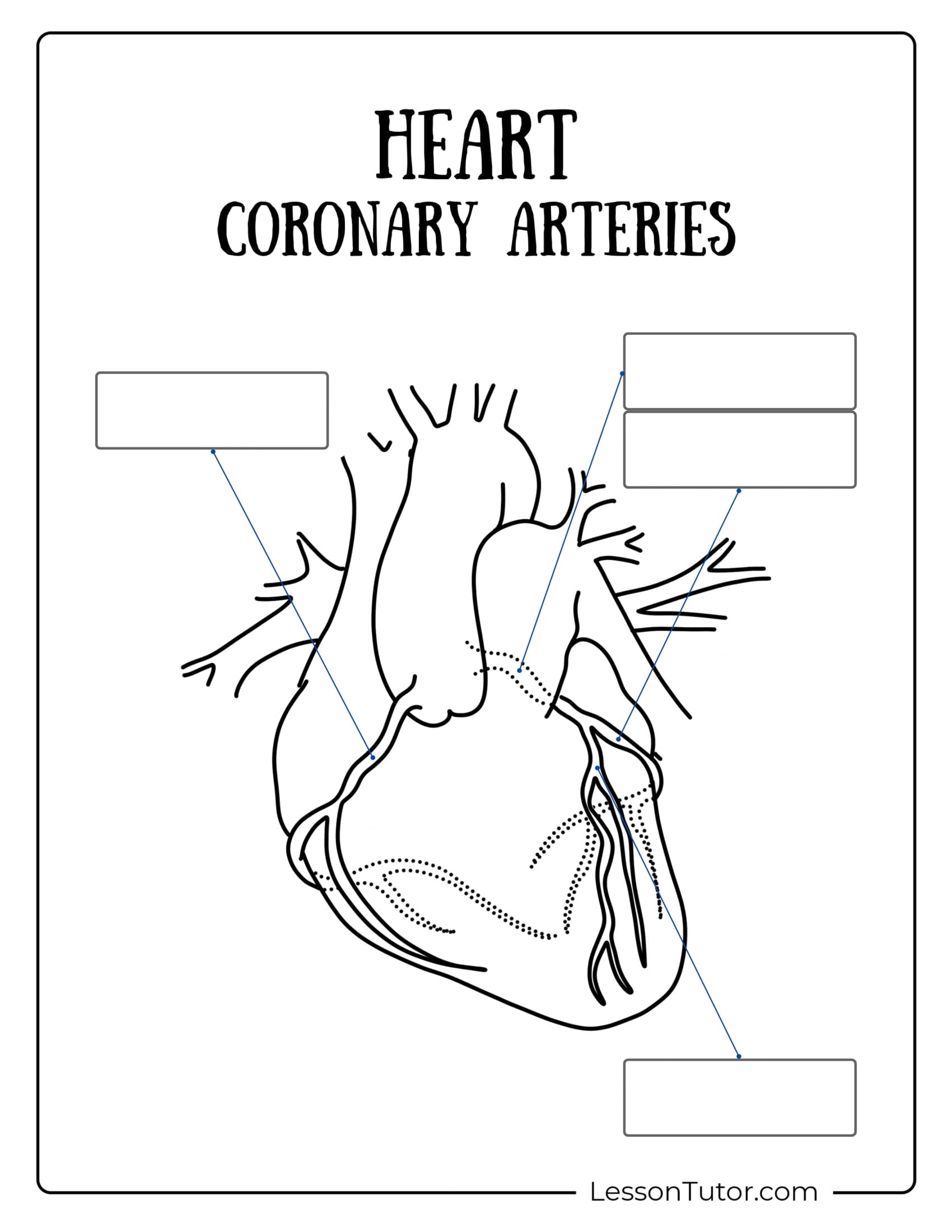
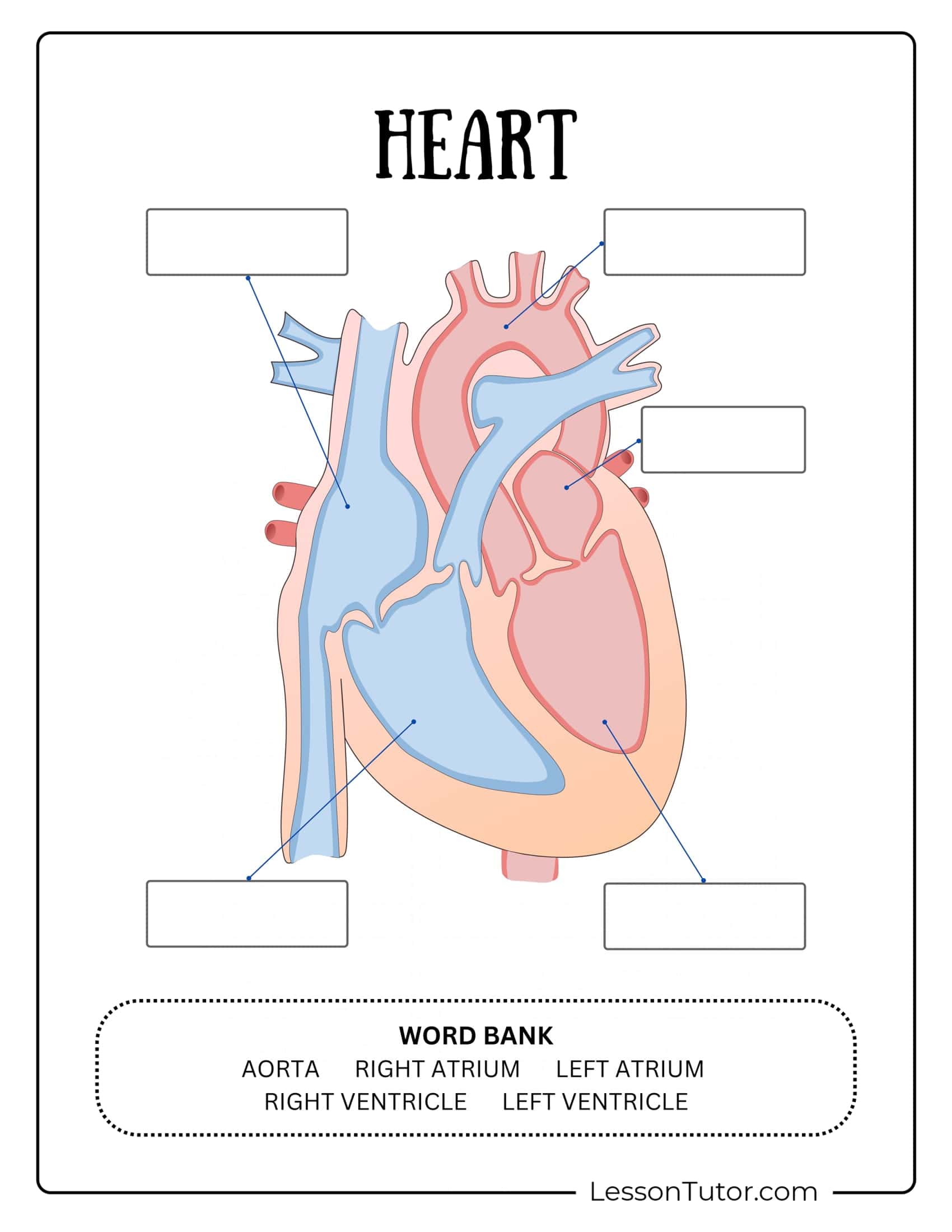
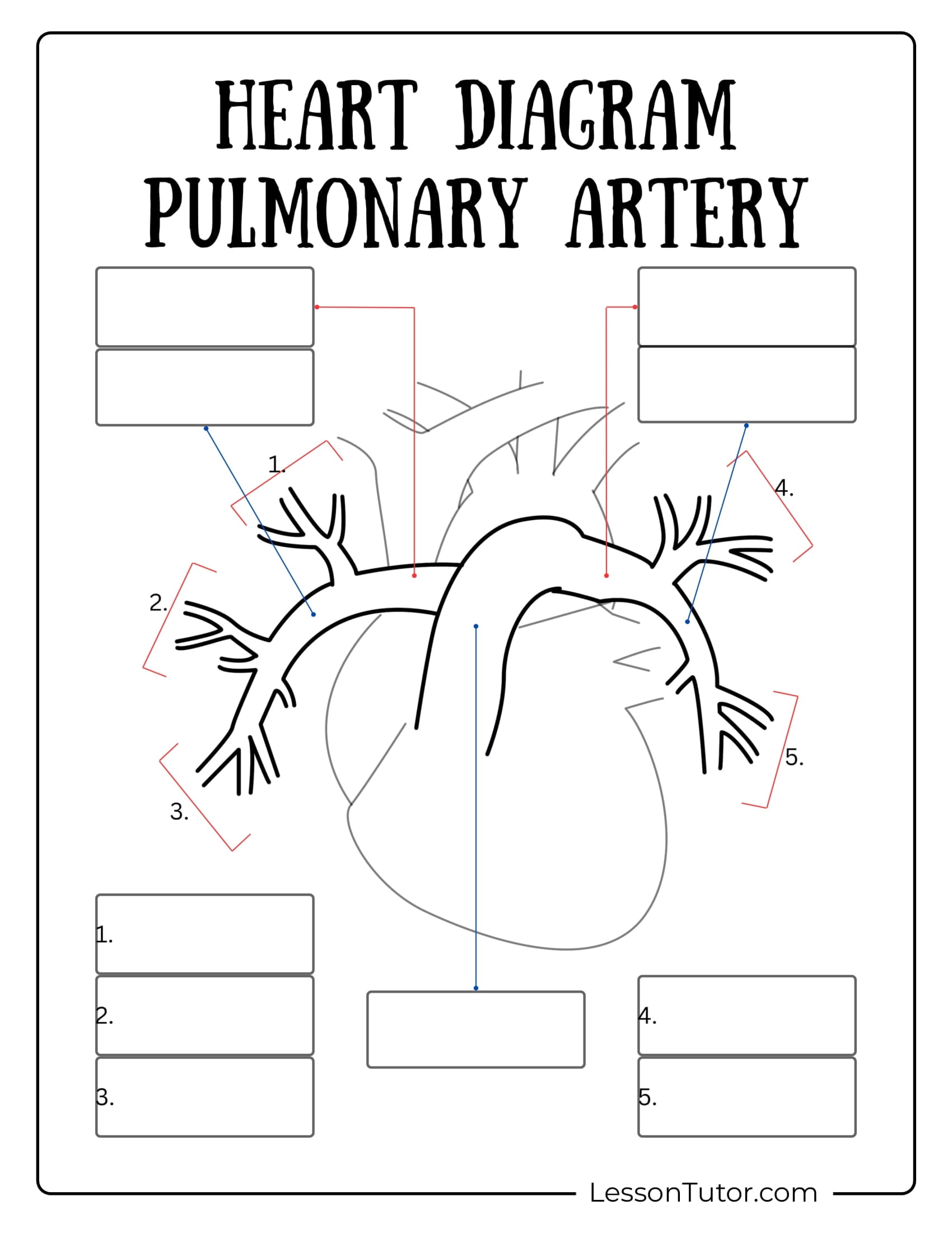
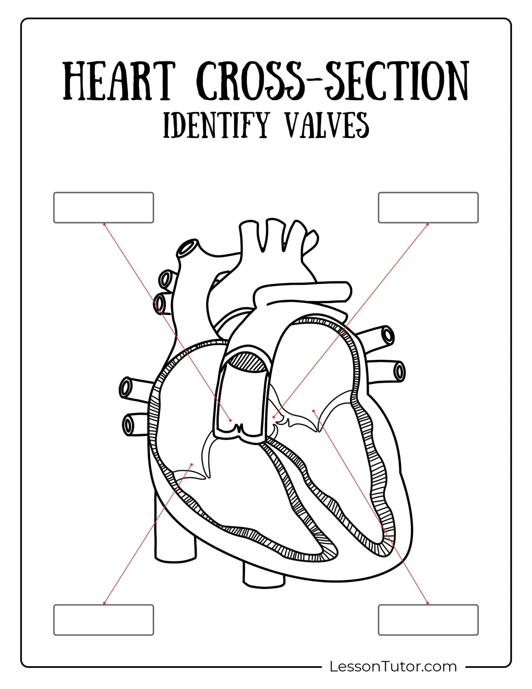
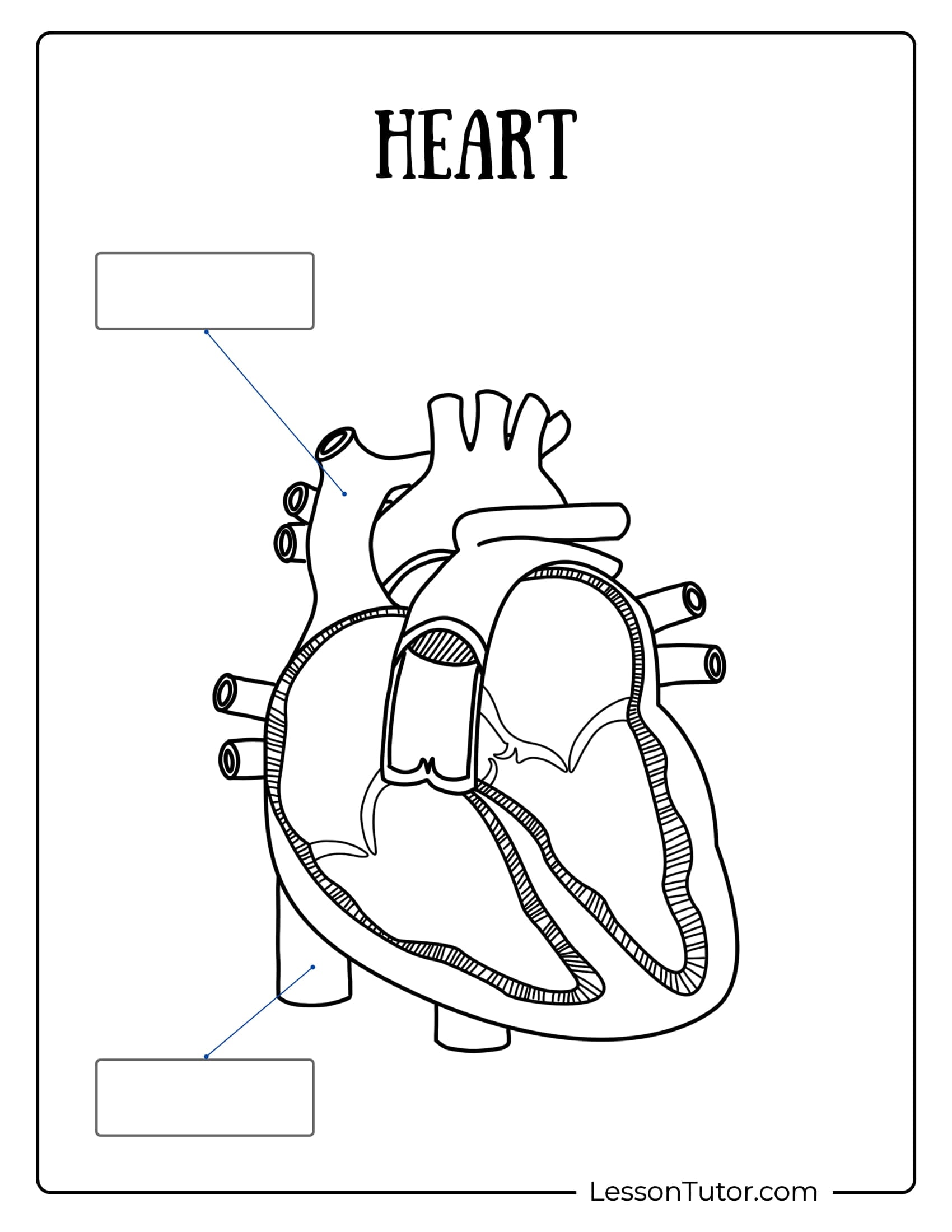
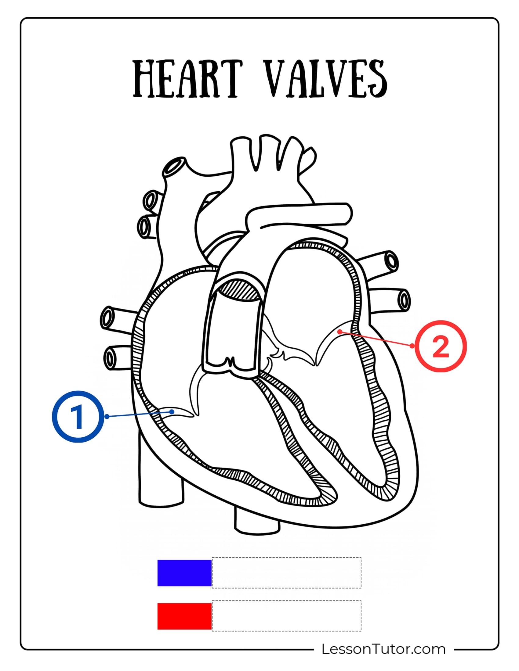
Immerse yourself in the fascinating anatomy of the human heart with this exclusive collection of 14 educational circulatory system worksheets designed for students, teachers, and curious minds eager to explore the wonders of the cardiovascular system! These detailed diagrams provide a hands-on learning experience, including a human heart with blank spaces for labeling chambers, a cross-section of the heart to identify valves, and a worksheet highlighting the aorta with blank labels. Whether you’re enhancing a lesson plan or diving deep into the fascinating anatomy of the heart, these worksheets offer hours of educational fun and discovery!
Step into the incredible world of the human heart, where science, anatomy, and discovery pulse with life! This exclusive collection of 14 meticulously designed, high-resolution circulatory system PDF worksheets, perfectly formatted for A4 paper, is perfect for curious learners and anatomy enthusiasts. Free to download and print, these engaging worksheets will guide you through the heart’s fascinating structure, from chambers to blood flow, ensuring hours of educational fun and discovery.
Craft Ideas To Do With Circulatory System Worksheets
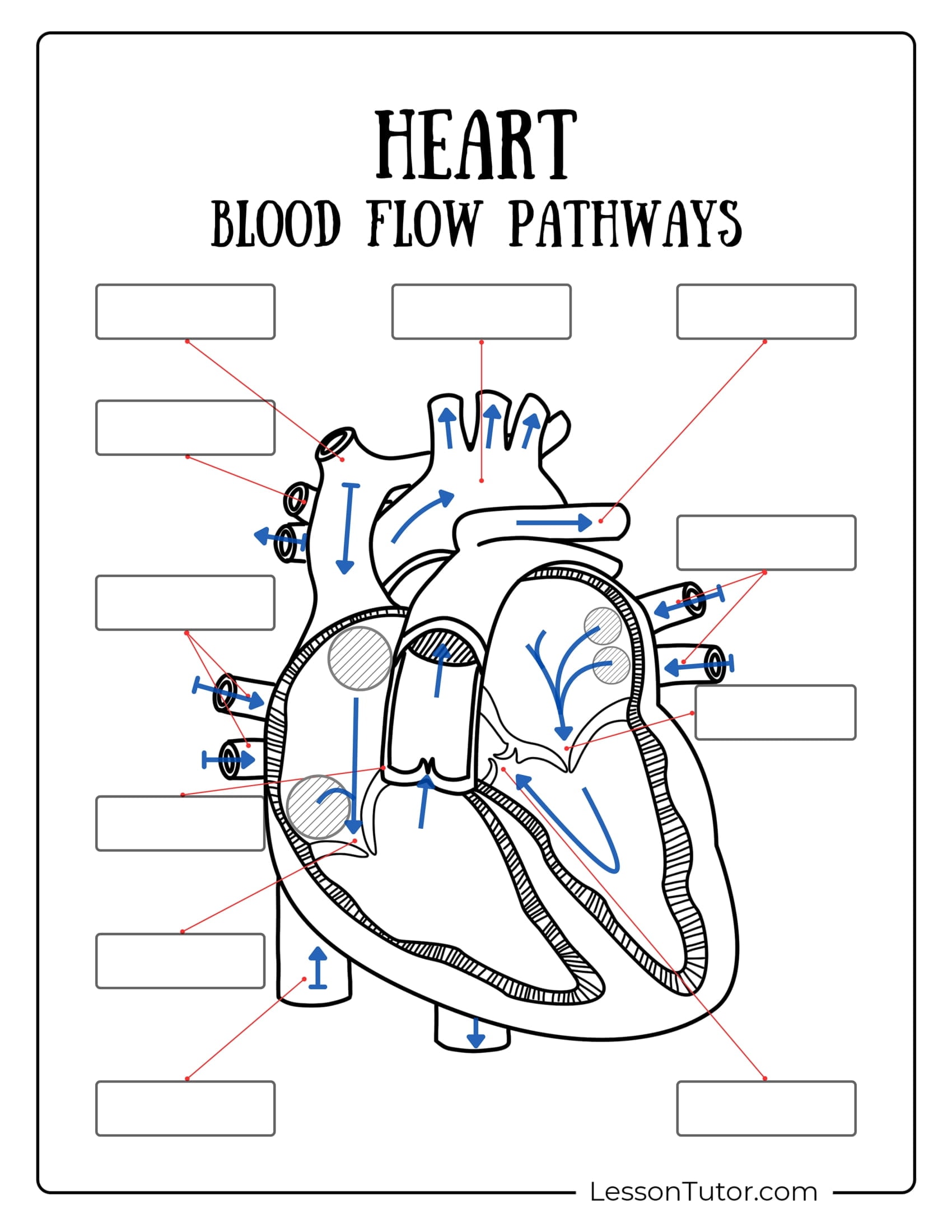
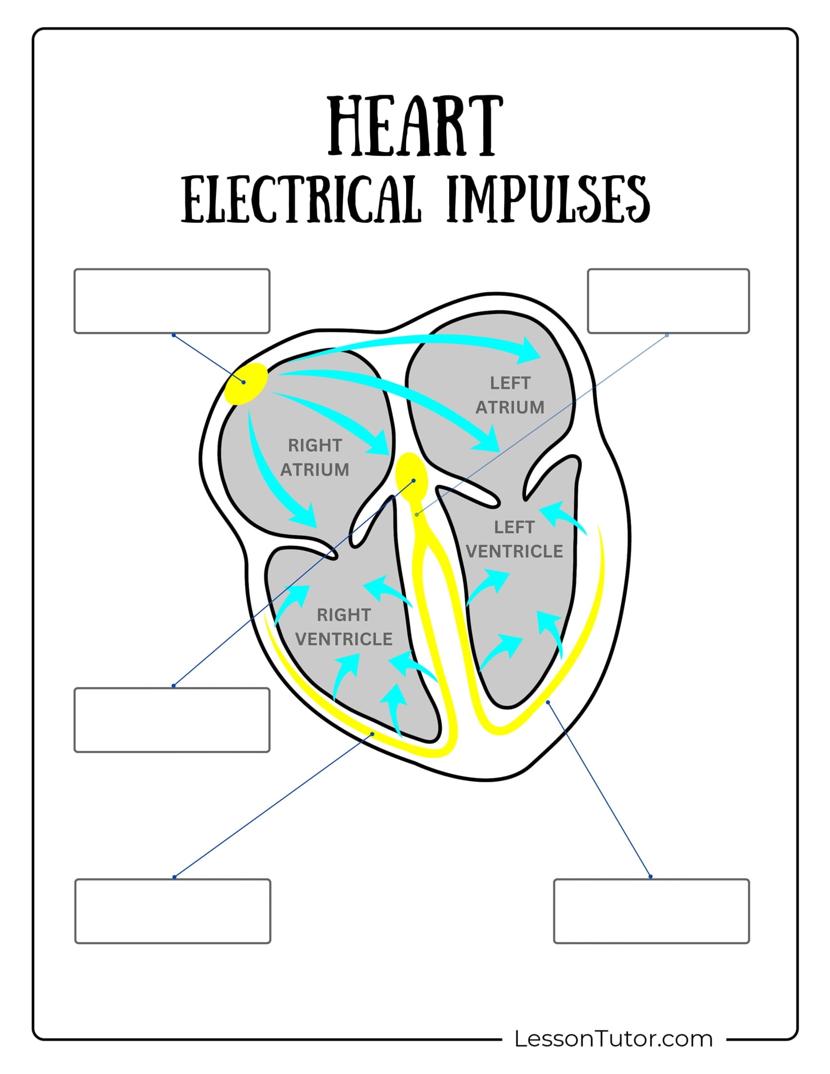
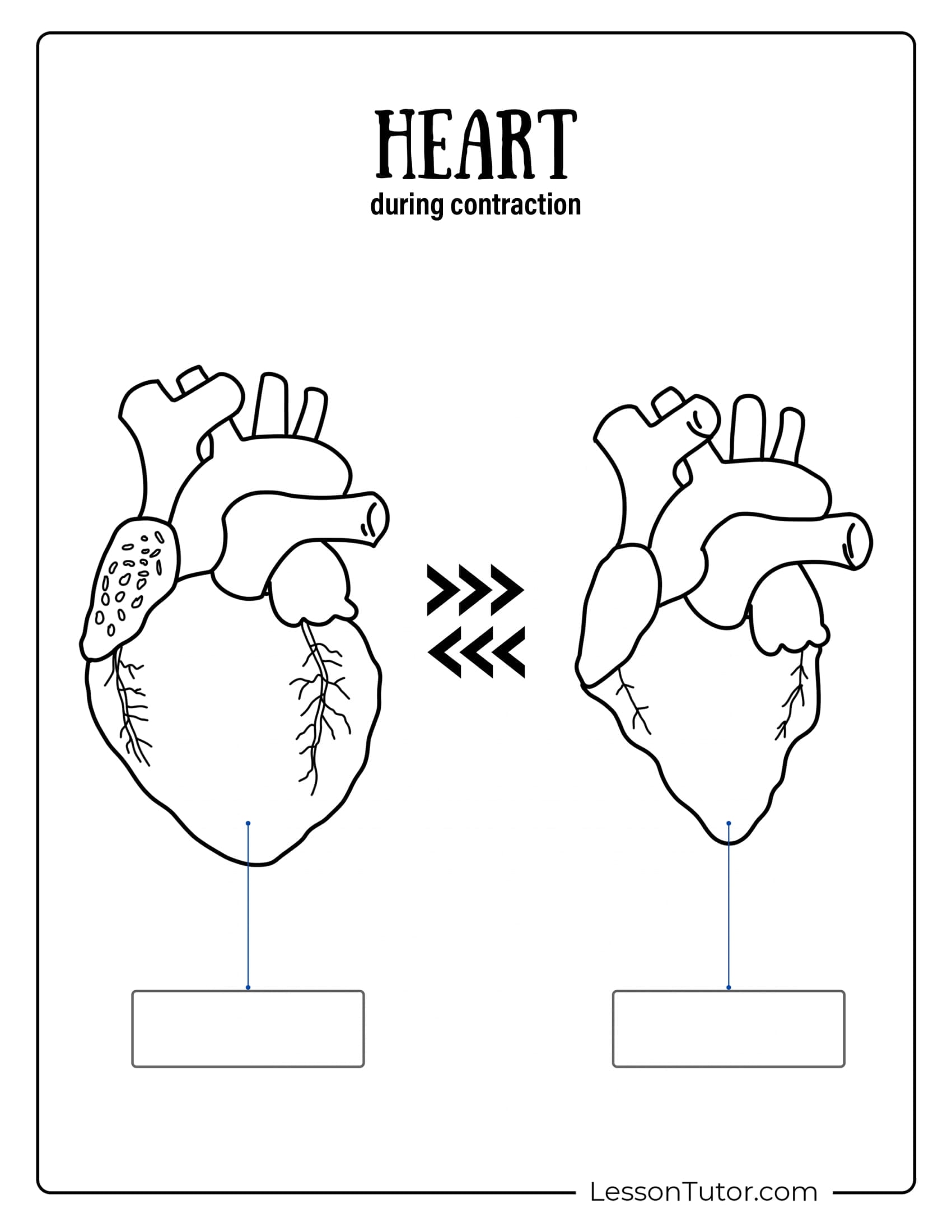
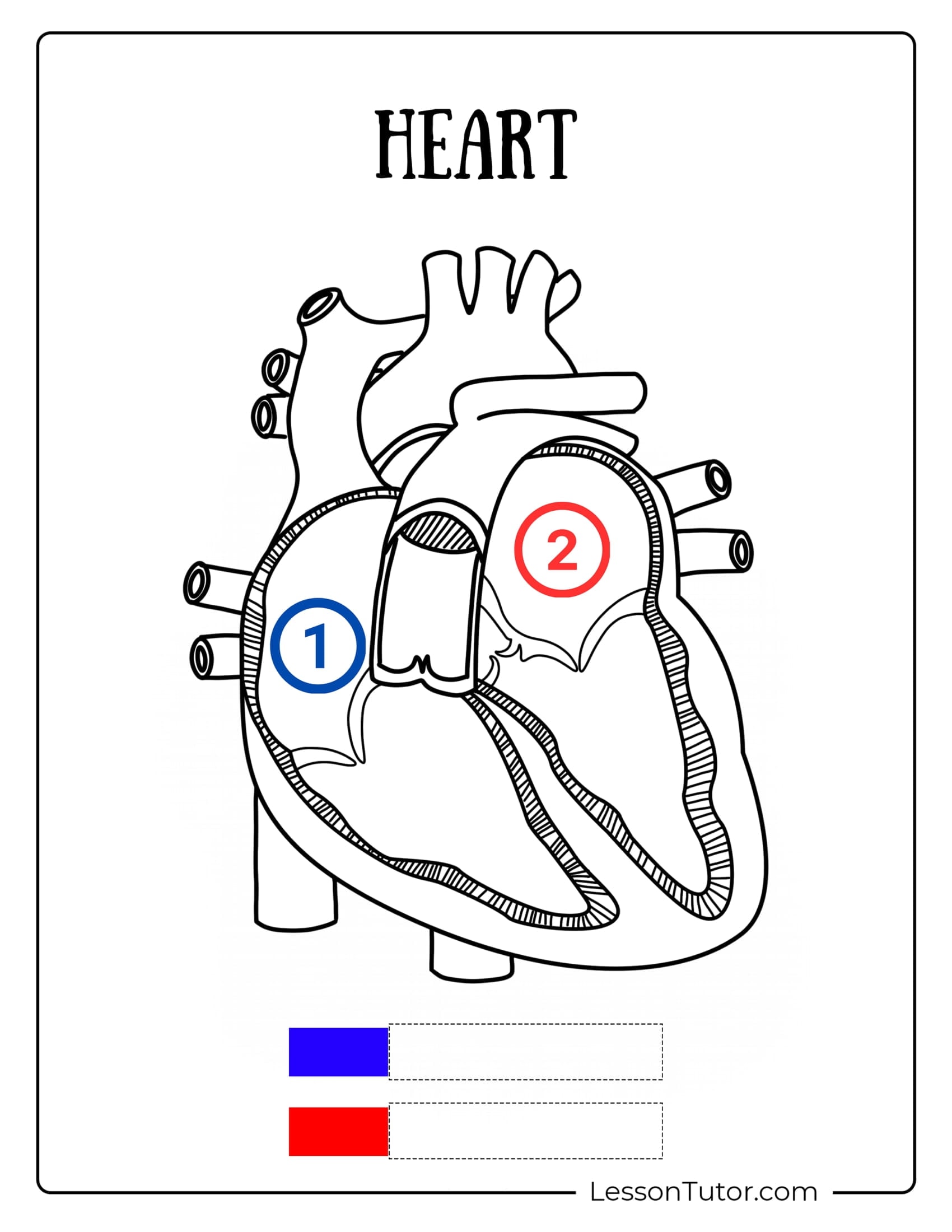
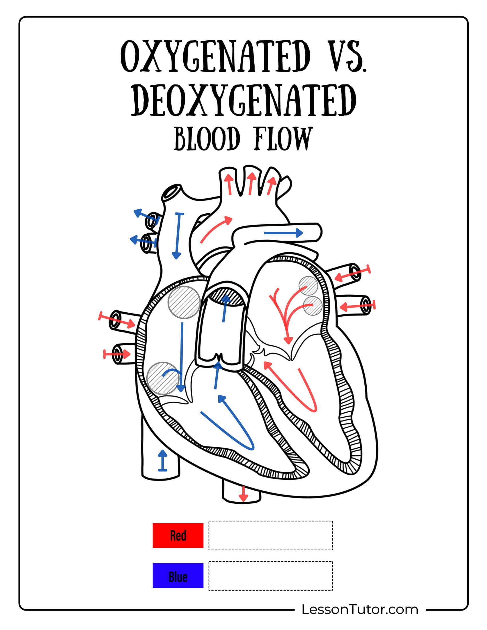
This set features diagrams illustrating oxygenated versus deoxygenated blood flow and the heart during contraction with blank spaces for labeling key components. Dive deeper into the science with a diagram showcasing the heart’s electrical impulses and numbered sections for identifying the left and right atriums. Whether you prefer brain-teasing puzzles, interactive quizzes, or detailed activity sheets, this collection of Respiratory System worksheets offers endless educational fun.
More Free Printable Worksheets
If you're looking for more related worksheet goodies that kids love, we think you'll particularly enjoy these worksheets collections: